Functional and Anatomical Imaging Research
Our group
FAIR is part of the Department of Information Engineering, University of Padova and it is leaded by Prof. Alessandra Bertoldo. The current members includes three postdoctoral fellows and four PhD students.
Our group
FAIR is part of the Department of Information Engineering, University of Padova and it is leaded by Prof. Alessandra Bertoldo. The current members includes three postdoctoral fellows and four PhD students.
Medical imaging, and the integration of imaging data with other information (either clinical, serological or -omical) is increasingly crucial for the understanding of disease progression, its early diagnosis and its monitoring. Moreover, it is becoming of fundamental importance for the study of pharmacodynamic and treatment efficacy in all districts where biopsy or sampling is infeasible.
Positron Emission Tomography (PET) and Magnetic Resonance Imaging (MRI) are powerful technologies that provide two- and three-dimensional images, which allow the evaluation of morphological changes and biological and physiological processes in vivo in humans. Quantitative analysis of PET, MRI data allows the estimation of important physiological parameters such as blood flow, glucose metabolism, oxygen consumption, receptor distribution.
Our aim
The aim of this research line is the study and development of new methodologies and/or models to estimate from anatomical and functional 2D/3D/4D images parameters characterizing peculiar dynamic processes.
Our aim
The aim of this research line is the study and development of new methodologies and/or models to estimate from anatomical and functional 2D/3D/4D images parameters characterizing peculiar dynamic processes.
In particular, PET research activity is primarily focused on the functional imaging of glucose metabolism in human skeletal muscle and lungs and on in vivo study of the interaction of tracers with specific receptor binding sites, such as the dopaminergic, adenosinergic and serotoninergic systems as well as quantification of TSPO inflammation tracers as [11C]PBR28. PET data are also studied in integration with genomic data to test mRNA gene expressions toward in vivo multimodal imaging data.
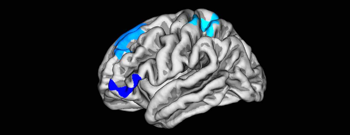
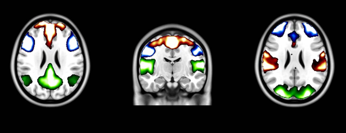
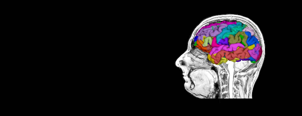
MRI research activity involves the quantitative analysis of two common methods for measuring perfusion with MRI, i.e. dynamic susceptibility contrast (DSC) and arterial spin labeling (ASL), the study of the diffusion anisotropy from diffusion tensor imaging (DTI) of the brain and skeletal muscle, the assessment of the brain iron accumulation in normal subjects and multiple sclerosis patients and the study of brain atrophy by using anatomical T1-weighted images. We also develop methods and models to derive activation maps from fMRI data sets, to study brain connectivity from resting state fMRI data and to integrate EEG signal and fMRI data.
Main Collaborations
-
Department of Neuroimaging, Institute of Psychiatry, Psychology and Neuroscience, King’s College London, UK (Paul Expert, Federico Turkheimer, Mattia Veronese)
-
Molecular Imaging Branch of the National Institute of Mental Health, Bethesda (USA) (Masahiro Fujita, Robert Innis, Sami Zoghbi)
-
PET centre of Guy’s and St Thomas’ National Health Service Trust, King’s College London, UK (Alexander Hammers, Colm McGinnity)
-
Neurological Institute, Houston Methodist Hospital, Houston (TX), USA (Paolo Zanotti Fregonara)
-
Columbia University, New York, NY (Todd Ogden, Francesca Zanderigo)
-
Policlinico Gemelli, Università Cattolica del Sacro Cuore, Roma, Italia (Maria Luisa Calcagni, Alessandro Giordano, Luca Indovina)
-
Università di Verona, Verona, Italia (Massimiliano Calabrese, Roberta Magliozzi, Francesca Pizzini)
-
Institute of Biomedical Engineering, University of Oxford, Oxford, UK (Michael Chappell)
-
Department of Neurosciences: SNPSRR, Università degli Studi di Padova, Padova, Italia (Maurizio Corbetta, Antonino Vallesi)
-
Leiden University Medical Center, Leiden, The Netherlands (Matthias Van Osch)
-
La nostra famiglia, Bosisio Parini, Lecco, Italia (Filippo Arrigoni, Denis Peruzzo)
-
Istituto Neurologico Casimiro Mondino, Pavia, Italia (Claudia Wheeler-Kingshott Gardini, Paolo Vitali)
-
Istituto Europeo di Oncologia, Milano Italia (Cristiano Crosta, Cristina Trovato)
-
Istituto Oncologico Veneto, Padova, Italia (Giorgio Battaglia, Stefano Realdon)
-
The University of New South Wales, Sydney, Australia (Rupert Leong, Hadis Mirzaei)
-
University of Dundee, Dundee, UK (Manuel Trucco)
-
Università Vita-Salute San Raffaele, Milano, Italia (Giulio Ferrari)
-
Jadavpur University, Jadavapur, India (Ananda Shankar Chowdhury)
-
Children’s National Hospital, Washington DC, USA (Marius G. Linguraru, Juan Cerrolaza)
-
University Medical Center , Utrecht, The Netherlands (Martin Froeling)


